Bone Health Risk Calculator
This calculator estimates your risk of developing osteoporosis after a bone marrow transplant based on key medical factors. Enter your information below to get personalized risk assessment.
Your Risk Assessment:
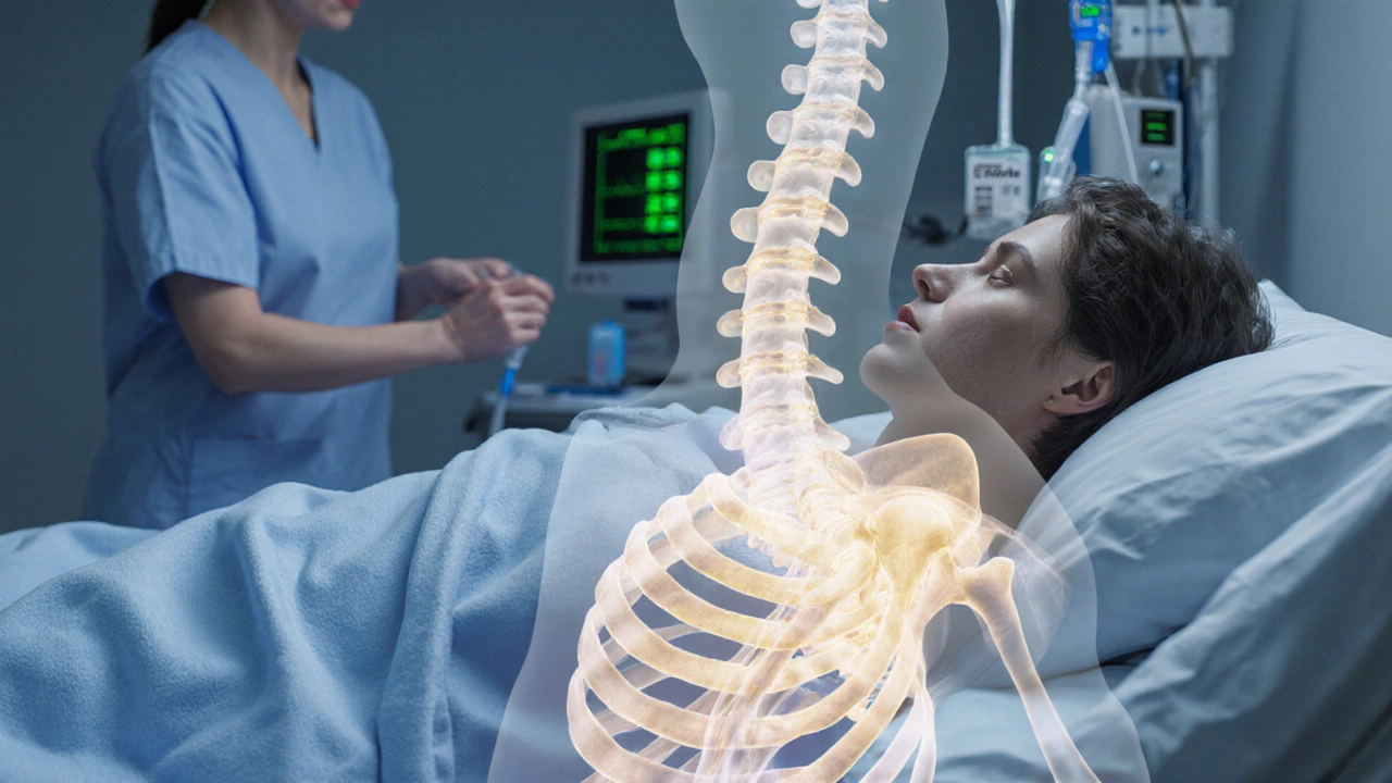
Facing a bone marrow transplant is already a lot to handle, but many patients don’t realize that a silent threat called osteoporosis can creep in during recovery. Understanding why this happens, how to spot early signs, and what you can do to protect your skeleton can make the difference between a smooth comeback and a painful setback.
Why Bone Health Takes a Hit After a Transplant
When a patient undergoes bone marrow transplant a procedure that replaces diseased or damaged marrow with healthy stem cells, the body goes through a cascade of stressors that tip the balance of bone remodeling.
- Glucocorticoid therapy: High‑dose steroids are standard for preventing graft‑versus‑host disease (GVHD) and managing inflammation. Steroids suppress osteoblast activity (bone‑building cells) while stimulating osteoclasts (bone‑resorbing cells), leading to rapid bone loss.
- Graft‑versus‑host disease: Chronic GVHD often requires prolonged immunosuppression, which further depletes calcium and vitamin D stores.
- Hormonal changes: The transplant can disrupt the endocrine axis, causing hypogonadism or thyroid dysfunction-both known culprits for weaker bones.
- Immobilization: Hospital stays, fatigue, and muscle weakness limit weight‑bearing activity, a key driver of bone strength.
These factors combine to push patients into a high‑risk category for osteoporosis a condition characterized by low bone mass and structural deterioration, increasing fracture risk-often within the first six months after the transplant.
Screening: How Doctors Detect Early Bone Loss
Early detection hinges on two tools:
- Dual‑energy X‑ray absorptiometry (DXA) the gold‑standard imaging test that measures bone mineral density (BMD) at the spine and hip. Baseline scans are usually performed before the transplant, with follow‑up scans at 3‑ to 6‑month intervals.
- Blood work for calcium, vitamin D, and markers of bone turnover (e.g., serum C‑telopeptide).
If the DXA T‑score drops below -1.0, clinicians typically start preventive measures; a score ≤ -2.5 confirms osteoporosis and triggers treatment.
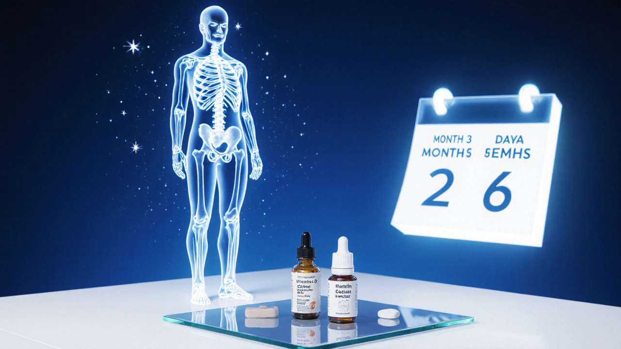
Nutrition and Supplements: The First Line of Defense
Even before medication enters the picture, a solid nutritional foundation can blunt bone loss.
- Calcium the mineral that provides structural support to bone tissue: Aim for 1,200mg daily from dairy, fortified plant milks, or supplements.
- Vitamin D a fat‑soluble vitamin that enhances calcium absorption in the gut: 800-1,000IU daily is common; levels below 30ng/mL warrant a higher dose.
- Protein intake of 1.2-1.5g/kg body weight supports muscle mass, which in turn loads bone.
- Limit caffeine (>300mg/day) and sodium (>2,300mg/day) as they can increase calcium excretion.
Regular monitoring of serum calcium and vitamin D every 1-2months during the steroid taper helps keep numbers in the therapeutic window.
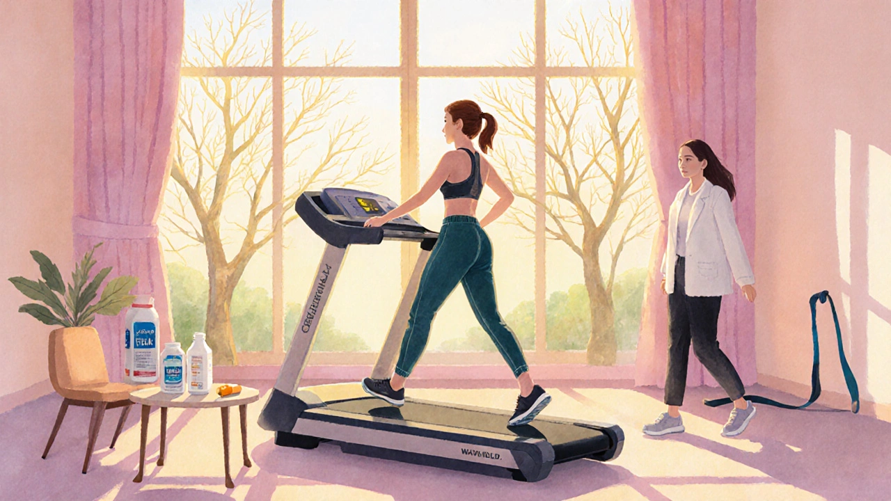
Exercise: Load the Skeleton Safely
Weight‑bearing and resistance activities send mechanical signals that tell osteoblasts to build, not break down.
- Start with low‑impact walking or stationary cycling for 15-20minutes, 3-5 times a week.
- Progress to resistance bands, body‑weight squats, and seated leg presses as tolerance improves.
- Balance training (e.g., single‑leg stands) reduces fall risk-a crucial factor since fractures often occur after a stumble.
Physiotherapists familiar with post‑transplant patients can tailor a program that respects fatigue levels and any line‑access wounds.
Medications: When Lifestyle Isn’t Enough
If DXA signals a significant decline, clinicians usually add pharmacologic therapy.
| Approach | Primary Benefit | Typical Start Time | Common Side Effects |
|---|---|---|---|
| Bisphosphonates (e.g., alendronate) | Inhibit osteoclast‑mediated bone resorption | Within 1-2months of steroid taper | Esophageal irritation, acute‑phase flu‑like symptoms |
| Denosumab (RANKL inhibitor) | Potent anti‑resorptive, works even when kidneys are compromised | When bisphosphonates are contraindicated | Hypocalcemia, skin infections |
| Lifestyle (nutrition + exercise) | Builds long‑term bone mass, improves overall health | From day1 of transplant prep | None when done correctly |
Oral bisphosphonates are the most frequently prescribed first‑line agents because they’re inexpensive and have decades of safety data. Intravenous options (zoledronic acid) are useful for patients who can’t swallow pills.
Denosumab is increasingly considered for those with chronic kidney disease post‑transplant, as it isn’t cleared renally.
Putting It All Together: A Sample Follow‑Up Timeline
- Pre‑transplant (4-6 weeks before): Baseline DXA, calcium & vitamin D labs, start calcium 1,200mg/day and vitamin D 800IU/day.
- Transplant admission: Ensure adequate hydration, avoid calcium‑binding drugs (e.g., high‑dose iron chelators).
- Weeks 0-4 post‑transplant: Continue supplements, begin gentle walking as tolerated, schedule first post‑transplant DXA at 3 months.
- Months 3-6: Review DXA results. If BMD loss >5% or T‑score ≤ -1.0, start oral bisphosphonate plus calcium/vit D boost (1,500IU). Add supervised resistance training.
- Month 12: Repeat DXA. If stable or improved, consider tapering bisphosphonate after 12-24 months, but maintain lifestyle measures indefinitely.
This timeline is flexible; patients with severe GVHD or prolonged steroid use may need earlier pharmacologic intervention.
Red Flags: When to Call Your Care Team
- Sudden, severe back or hip pain without injury (possible vertebral fracture).
- Unexpected loss of height or a noticeable curvature in the spine.
- Signs of low calcium: tingling in fingers, muscle cramps, or heart rhythm irregularities.
- Any new medication that interferes with calcium absorption (e.g., certain anticonvulsants).
Prompt evaluation can prevent a small fracture from becoming a major setback that delays further transplant milestones.
Frequently Asked Questions
How soon after a bone marrow transplant can osteoporosis develop?
Bone loss can begin within the first few weeks, especially if high‑dose steroids are used. Most clinicians see measurable changes on DXA by 3months.
Are bisphosphonates safe for transplant patients?
Yes, when taken as directed they are well‑tolerated. Oral forms require staying upright for 30minutes after each dose to avoid esophageal irritation. Intravenous zoledronic acid is an option for those with swallowing difficulties.
Can I rely only on calcium and vitamin D without medication?
Supplements are essential but rarely enough on their own when steroids are involved. They work best as part of a combined strategy that includes exercise and, when needed, anti‑resorptive drugs.
What type of exercise is safest during the early recovery phase?
Low‑impact, weight‑bearing activities such as short walks, stationary cycling, and gentle resistance band work are safest. Avoid heavy lifting or high‑impact sports until cleared by your transplant team.
How often should I have a DXA scan after transplant?
A baseline scan pre‑transplant, then a follow‑up at 3months. If bone loss is detected, repeat every 6months until stability, after which annual scans are typical.
Staying proactive about bone health doesn’t have to feel overwhelming. With the right mix of nutrition, movement, monitoring, and-when needed-medication, most transplant recipients keep their skeleton strong enough to get back to the activities they love.

Brian Davis
October 6, 2025 AT 15:44Undergoing a bone marrow transplant initiates a cascade of physiological stressors that, if unmitigated, can precipitously erode skeletal integrity; the interplay between high‑dose glucocorticoids, prolonged immobilization, and endocrine disruption forms a triad of osteopenic risk.
First, glucocorticoids suppress osteoblast differentiation while simultaneously enhancing osteoclast activity, a mechanism well documented in endocrinology literature.
Second, the requisite period of reduced weight‑bearing activity diminishes the mechanical loading essential for bone remodeling, thereby accelerating loss of trabecular density.
Third, transplant‑related hormonal perturbations, such as hypogonadism and altered thyroid function, further compromise calcium homeostasis.
Evidence suggests that measurable declines in bone mineral density can be observed as early as six weeks post‑transplant, underscoring the necessity of early screening.
A baseline dual‑energy X‑ray absorptiometry (DXA) scan prior to conditioning provides a reference point for subsequent monitoring.
Subsequent DXA assessments at three‑month intervals allow clinicians to quantify the rate of bone loss and to tailor therapeutic interventions accordingly.
Calcium supplementation, targeting an intake of approximately 1,200 mg per day, should be initiated pre‑emptively, supplemented by vitamin D in the range of 800–1,000 IU daily, with adjustments based on serum 25‑hydroxyvitamin D levels.
Protein intake, calibrated at 1.2–1.5 g per kilogram of body weight, supports muscle mass which indirectly benefits bone through mechanotransduction.
Weight‑bearing exercise, even in modest form such as short indoor walks or stationary cycling, re‑establishes osteogenic stimulus without overwhelming the patient’s limited energy reserves.
When DXA reveals a T‑score ≤ −1.0, pharmacologic therapy is generally indicated; oral bisphosphonates remain first‑line due to extensive safety data, whereas intravenous zoledronic acid offers an alternative for those with dysphagia.
In cases of renal insufficiency, denosumab provides a viable anti‑resorptive option, though clinicians must vigilantly monitor for hypocalcemia.
Regular monitoring of serum calcium and phosphate, alongside vitamin D, every one to two months during steroid tapering, aids in preventing secondary hyperparathyroidism.
Patient education concerning fracture red‑flags-such as sudden back pain, loss of height, or spinal curvature-facilitates prompt medical attention and mitigates the risk of catastrophic skeletal events.
Ultimately, a multidisciplinary approach encompassing hematology, endocrinology, nutrition, and physiotherapy optimizes bone health outcomes and enhances overall quality of life post‑transplant.
jenni williams
October 16, 2025 AT 09:04hey there, just wanted to say you’re not alone in this journey 😊 keep up with your calcium and vitamin D, even when it feels like a lot, you’re doing great.
Kevin Galligan
October 26, 2025 AT 02:24Wow, that was a dissertation in a paragraph-thanks for the PhD crash course, now I know exactly why my knees creak after chemo 😂. If only the steroids came with a side‑effect disclaimer that said “you’ll also lose bone density”.
Dileep Jha
November 4, 2025 AT 18:44While the layperson’s hyperbole is entertaining, the pathophysiological substrate warrants a more nuanced exposition; the osteoclastic surge is mediated via RANK‑L upregulation secondary to cytokine cascades, not merely “steroid side‑effects”. Moreover, the biomechanical disuse osteopenia aligns with Wolff’s law deviations, necessitating mechanotransductive interventions.
Michael Dennis
November 14, 2025 AT 12:04The article presents a comprehensive overview, yet it could benefit from a more succinct executive summary; readers may appreciate a distilled bullet‑point section highlighting key interventions without wading through extensive narrative.
Blair Robertshaw
November 24, 2025 AT 05:24yeah, that “executive summary” thing is just fluff – who has time to read bullet points when the real problem is that doctors still ignore early DXA scans, lol.
Alec Maley
December 3, 2025 AT 22:44Totally get that the info can feel overwhelming; just take it one step at a time-start with the supplements and a short walk each day, and the rest will follow when you’re ready.
Navjot Ghotra
December 13, 2025 AT 16:04Sounds good
Claus Rossler
December 23, 2025 AT 09:24One must question the prevailing orthodoxy that universal bisphosphonate prophylaxis is the panacea; individualized risk stratification, perhaps integrating genetic polymorphisms of the VDR gene, may render a more sophisticated therapeutic algorithm.
chris mattox
January 2, 2026 AT 02:44Hey folks, think of your bones like a vibrant garden-water them with calcium, give them sunshine with vitamin D, and prune away the sedentary habits; together we can cultivate resilience and bloom beyond the transplant.
Jackson Whicker
January 11, 2026 AT 20:04Behold the skeletal symphony, each mineral note trembling under the conductor’s baton of endocrine whispers; neglect this orchestra and you shall hear the cacophony of fractures echoing through the corridors of recovery.
Audrin De Waal
January 21, 2026 AT 13:24Our bodies are the fortresses of our nation’s spirit-let’s not let foreign steroids pillage our bone heritage, stand tall and protect your marrow legacy.
parag mandle
January 31, 2026 AT 06:44Listen, the first 90 days post‑transplant are a crucible; by instituting a regimented regimen of calcium 1,200 mg, vitamin D 1,000 IU, and supervised resistance training, you forge an indomitable skeletal framework that will outlast the immunosuppressive tempest.
Shivali Dixit Saxena
February 10, 2026 AT 00:04Start calcium 1200 mg daily; boost vitamin D to 2000 IU if deficient; add weight‑bearing exercise 3×/week; re‑check DXA at 3‑month intervals; monitor labs bi‑monthly; adjust therapy as needed.
Sayam Masood
February 19, 2026 AT 17:24In the grand tapestry of recovery, the threads of bone health intertwine with hope; neglecting them unravels the fabric of resilience, so we must weave diligence into every clinic visit.
Jason Montgomery
March 1, 2026 AT 10:44Yo, keep that momentum-track your supplements, set a reminder for that weekly walk, and remember: consistency beats intensity every time.
Wade Developer
March 11, 2026 AT 04:04From a clinical perspective, the integration of pharmacologic and lifestyle interventions reflects a holistic paradigm; philosophically, it underscores the principle that preventive care is the most ethical stewardship of patient autonomy.
Sandra Perkins
March 20, 2026 AT 21:24Oh great, another “must‑do” list-because we all have endless free time, right?
rama andika
March 30, 2026 AT 15:44Sure, the “official” guidelines are fine-until you realize they’re penned by a cabal of pharma giants who profit from every bisphosphonate pill you swallow, all while whispering that your bones will crumble without them 😒.